en
Synovial entrapment in alloplastic temporomandibular joint replacement
/social-network-service/media/default/6809/fbab08d0.jpg)
15
12 min read
22 April 2025
Abstract.
Complications of alloplastic temporomandibular joint (TMJ) prostheses can lead to stress and anxiety for the patient and the surgical team, and prosthesis substitution is sometimes required. The aim of this case report is to describe the surgical finding of synovial entrapment with interposed fibrosis in a postoperative alloplastic TMJ revision, managed effectively with adequate surgical debridement. The authors believe that synovial entrapment needs to be considered as a possible postoperative complication of total joint replacement when no clear symptoms of infection, metal hypersensitivity, osteolysis, or heterotopic bone formation are present. The implications of synovial entrapment in TMJ alloplastic replacement remains relatively unpredictable and poorly understood.
The number of alloplastic temporomandibular joint replacement (TMJR) procedures is expected to increase 58% by the year 2030. In a survey of 4638 TMJR procedures, the incidence of TMJR surgical revision was 3% and the incidence of replacement was 4.9%. Despite the relatively low incidence of TMJR revision, from a clinical and economic perspective, the consequences are relevant. Therefore, it is important to report all possible causes of TMJR complications with the main goal of progressing medical knowledge, increasing predictability, and consequently improving patient quality of life.
The most common complications of TMJR are periprosthetic joint infection, heterotopic bone formation, dislocation of the condylar component, continued post-TMJR pain, material hypersensitivity, neuroma formation, screw loosening, fracture, and synovial entrapment syndrome. Synovial entrapment is a relatively new concept in TMJR; however, in the orthopaedic field, synovial-like fibrous membranes were first reported after total hip and knee replacement in 1986 and 1990, respectively. Subsequently, Jerosch and Schroder described the development of intra-articular plicae as a cause of pain in knee arthroplasty. Synovial entrapment syndrome in TMJR was first reported in 2010 by Westermark et al. for Biomet (Biomet Microfixation, Jacksonville, FL, USA) and TMJ Concepts prostheses (TMJ Concepts, Ventura, CA, USA). However, current understanding of the signs and symptoms of synovial entrapment as a complication of TMJR is still insufficient.
This article describes a case of symptomatic postoperative TMJR, managed effectively with adequate surgical debridement. The surgical finding of synovial entrapment with interposed fibrosis was determined to be a possible cause of the symptoms.
Case report
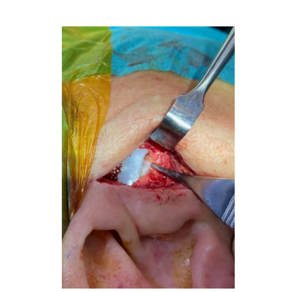
A healthy 35-year-old woman was referred to our department after several failed TMJ surgeries. The patient had persistent pain and limited mouth opening. Imaging exams revealed a right fibrous ankylosis. She underwent a unilateral custom TMJR (TMJ Concepts). At 2 years and 5 months after TMJR, the patient demonstrated new episodes of pain at the operated site (score 7/10 on a visual analogue scale (VAS)), with intermittent swelling and progressive mouth limitation (reduced from 37 mm to 16 mm). No improvement occurred with antibiotics, non-steroidal anti-inflammatory drugs (NSAIDs), or physiotherapy. Changes were not observed on computed tomography scan and no infection criteria were present. At first, the authors hypothesized a diagnosis of metal hypersensitivity. However, lymphocyte transformation testing is not available in Portugal, so this hypothesis was not confimed. With no clear diagnosis, it was decided that additional open surgery should be performed to debride the joint area. Considerable fibrosis resembling synovial tissue was noted surrounding the joint area, with part of the tissue showing an inflammatory appearance. Fibrous/synovial tissue was also observed between the alloplastic condyle and fossa (Fig. 1). This interposed tissue mimicking a joint disc in an alloplastic reconstruction was a surprising and unexpected finding.
All tissue was excised and carefully electrocoagulated, with special care to avoid scuffing the alloplastic components of the TMJR device. The joint area was scrubbed with iodine solution and irrigated with vancomycin and gentamicin. The total surgery time was 28 minutes. Histopathological assessment demonstrated the presence of synovial tissue with low inflammatory infiltration (Fig. 2), suggesting synovial entrapment. At the 6-month postoperative follow-up, the patient was pain-free (VAS pain score of 0/10), with a maximum mouth opening of 36 mm.

Discussion
Over the last few years, several reports have addressed the indications for revision and replacement of the alloplastic TMJ. The most common extrinsic causes are infection, wear and fracture of the prosthesis, allergy to the prosthesis, foreign body reaction, heterotopic bone formation, dislocation of the condyle component, osteolysis, neuroma formation, and synovial entrapment syndrome. Some intrinsic aetiologies have also been described: chronic centrally mediated pain, persistent myofascial/muscular pain, complex regional pain syndrome I, temporalis tendonitis, coronoid impingement, Frey syndrome, and integrin formation.
In most reports, the implications of synovial entrapment in TMJR remain relatively unnoticed. This phenomenon has been the subject of debate in recent years and its association with symptoms is controversial. Hardaker et al. described thickened and fibrotic synovial plicae with some inelasticity, consequently leading to synovitis, chondral damage, and pain. Hypotheses related to synovial entrapment in alloplastic devices include: (1) a response of the mesenchymal tissues surrounding the prosthesis; (2) the chemical and physical composition of the replacement elements; and (3) the presence of molecules involved
in inflammatory processes, such as interleukin-1b and interleukin-6, tumour necrosis factor-alpha, transforming growth factor-beta, and prostaglandin-E2, as shown in some biochemical studies in synovial-like tissue. The release of these molecules can be related to mechanical stress.
This phenomenon has also been confirmed in the TMJ, related to internal derangement. It was described for the first time in TMJ alloplastic devices by Westermark et al. as a dense, fibrous connective tissue without signal of inflammatory cells or foreign body reactions and synovial-like tissue between the capsule and disc observed in TMJ prosthesis revision (Biomet Microfixation and TMJ Concepts). Also, Monje et al. reported a case of synovial metaplasia with a temporary silicone implant. Recently, Davis et al., reported a case series of nine patients with failed TMJ prostheses, managed with TMJ arthroscopy. They verified the presence of a pseudocapsule between the fossa and condylar portion, and biopsy results showed synovium, fibrous connective tissue, degenerated fibrocartilage, and focal dystrophic calcifications, with negative results for pathogens. Similar to TMJR, other orthopaedic surgeons have described fibrous plicae after alloplastic knee replacement in 26 patients in five different zones, for which arthroscopic resection successfully resolved the patients’ symptoms. Arthroscopic interventions for problematic alloplastic TMJR should be restricted to experienced arthroscopic surgeons to avoid damage the alloplastic joint components, and an open approach would be more suitable for the less experienced arthroscopist.
It appears that the association of synovial plicae with symptoms and clinical findings continues to be controversial. However, the present report reinforces the possible implications of synovial entrapment with an interpositional fibrous pseudodisc between the alloplastic condyle and fossa as a TMJR complication.
A limitation of this study was that cultures of the tissues to exclude the hypothesis of low-grade biofilm, as previously described by Gruber et al., were not performed. The authors believe that synovial entrapment needs to be considered as a possible postoperative complication of TMJR when no clear symptoms of infection, metal hypersensitivity, osteolysis, or heterotopic bone formation are present. If this pathological entity is suspected, debridement as a surgical revision can provide clinical improvement, either by arthroscopy or open surgery, depending on the surgeon experience. Future studies in this field may help to develop easier ways to diagnose this phenomenon in alloplastic joints as it remains relatively unpredictable and poorly understood.
Authors: D. F. Ângelo, H. J. Cardoso, D. Sanz
References:
- Onoriobe U, Miloro M, Sukotjo C, Mercuri LG, Lotesto A, Eke R. How many temporomandibular joint total joint alloplastic implants will be placed in the United States in 2030? J Oral Maxillofac Surg 2016;74:1531–8.
- Amarista FJ, Mercuri LG, Perez D. Temporomandibular joint prosthesis revision and/or replacement survey and review of the literature. J Oral Maxillofac Surg 2020;78:1692–703.
- Mercuri LG. Complications associated with TMJ TJR: management and prevention. In: Mercuri L, editor. Temporomandibular joint total joint replacement – TMJ TJR: a comprehensive reference for researchers, mate rials scientists, and surgeons. Cham, Switzerland: Springer; 2016 . p. 187–226. http://dx.doi.org/10.1007/978-3-319-21389- 7_8.
- Goldring SR, Jasty M, Roelke MS, Rourke CM, Bringhurst FR, Harris WH. Formation of a synovial-like membrane at the bone– cement interface. Its role in bone resorption and implant loosening after total hip replacement. Arthritis Rheum 1986;29:836–42.
- Thorpe CD, Bocell JR, Tullos HS. Intra-articular fibrous bands. Patellar complications after total knee replacement. J Bone Joint Surg Am 1990;72:811–4.
- Jerosch J, Schroder M. Clinical symptoms caused by intra-articular fibrous plicae after knee replacement. Arthroscopic diagnosis and therapy. Arch Orthop Trauma Surg 1996;115:195–8.
- Westermark A, Leiggener C, Aagaard E, Lindskog S. Histological findings in soft tissues around temporomandibular joint prostheses after up to eight years of function. Int J Oral Maxillofac Surg 2011;40:18–25.
- Gakhal MK, Gupta B, Sidebottom AJ. Analysis of outcomes after revision replacement of failed total temporomandibular joint prostheses. Br J Oral Maxillofac Surg 2020;58:220–4.
- Hardaker WT, Whipple TL, Bassett 3rd FH. Diagnosis and treatment of the plica syndrome of the knee. J Bone Joint Surg Am 1980;62:221–5.
- Edwards JC, Sedgwick AD, Willoughby DA. The formation of a structure with the features of synovial lining by subcutaneous injection of air: an in vivo tissue culture system. J Pathol 1981;134:147–56.
- Murray PD, Drachman DB. The role of movement in the development of joints and related structures: the head and neck in the chick embryo. J Embryol Exp Morphol 1969;22:349–71.
- Raso DS, Crymes LW, Metcalf JS. Histological assessment of fifty breast capsules from smooth and textured augmentation and reconstruction mammoplasty prostheses with emphasis on the role of synovial metaplasia. Mod Pathol 1994;7:310–6.
- Monje F, Mercuri L, Villanueva-Alcojol L, de Mera JJ. Synovial metaplasia found in tissue encapsulating a silicone spacer during 2-staged temporomandibular joint replacement for ankylosis. J Oral Maxillofac Surg 2012;70:2290–8.
- Konttinen YT, Waris V, Xu JW, Jiranek WA, Sorsa T, Virtanen I, Santavirta S. Transforming growth factor-beta 1 and 2 in the synovial-like interface membrane between implant and bone in loosening of total hip arthroplasty. J Rheumatol 1997;24:694–701.
- Bosetti M, Massè A, Navone R, Cannas M. Biochemical and histological evaluation of human synovial-like membrane around failed total hip replacement prostheses during in vitro mechanical loading. J Mater Sci Mater Med 2001;12:693–8.
- Murakami K, Moses JJ, Israel H, McCain JP. Synovial plica, lateral impingement and intra-articular impingement/entrapment pathologies of the temporomandibular joint. Front Oral Maxillofac Med 2020;2:24.
- Murakami K, Hori S, Yamaguchi Y, Mercuri LG, Harayama N, Maruo S, Takahashi T. Synovial plicae and temporomandibular joint disorders: surgical findings. J Oral Maxillofac Surg 2015;73:827–33.
- Davis CM, Hakim M, Choi DD, Behrman DA, Israel H, McCain JP. Early clinical outcomes of arthroscopic management of the failing alloplastic temporomandibular joint prosthesis. J Oral Maxillofac Surg 2020;78:903–7.
- Gruber EA, McCullough J, Sidebottom AJ. Medium-term outcomes and complications after total replacement of the temporomandibular joint. Prospective outcome analysis